The Ink of Life: How 3D Bioprinting is Quietly Rewriting the Future of Medicine
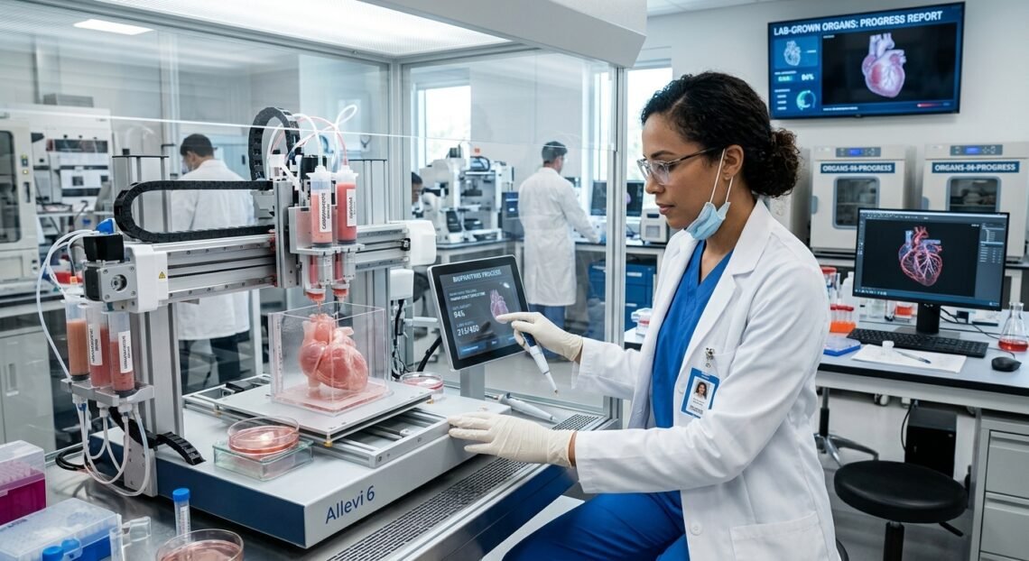
If you spent any time reading sci-fi a few decades ago, the idea of printing a fully functioning human organ on demand sounded as realistic as warp drives or teleporters. Yet right now, in quiet research labs across the world, scientists are loading specialized printers not with plastic or ink, but with living human cells.
We are standing on the edge of a massive shift in medicine. The global organ donor shortage has long been a devastating crisis—tens of thousands of people sit on transplant waiting lists, waiting for a compatible donor while their time slowly runs out. 3D bioprinting and lab-grown organs aren’t just promising to shorten that line; they are aiming to wipe it out entirely.
What Does Bioprinting Actually Look Like?
When people hear “3D printing,” they usually imagine a nozzle melting a spool of plastic to build a toy or a spare car part. Bioprinting works on a similar mechanical principle, but the materials involved are infinitely more fragile.
Instead of plastic, bioprinters use something called bioink. Bioink is typically a hydrogel—a squishy, water-rich compound made of natural or synthetic polymers—mixed with living cells. The gel acts as a temporary scaffold, keeping the cells in place and providing them with nutrients so they don’t collapse into a soup while the printer lays down layer after microscopic layer.
The process generally follows three main stages:
- The Blueprint: Doctors use high-resolution MRI or CT scans to map out the exact 3D geometry of a patient’s target tissue or organ.
- The Print Run:Using a robotic printhead, the bioprinter deposits bioink according to the digital map, building biological structures with pinpoint precision.
- Maturation in a Bioreactor: Once printed, the construct isn’t instantly ready to be implanted. It goes into a specialized chamber called a bioreactor, which mimics the body’s natural environment—providing warmth, oxygen, nutrient flow, and mechanical stress so the cells grow together and learn to function as a unified tissue.
Where the Field Stands Today
It’s easy to get swept up in the hype, but it helps to separate what we can do today from what is still a few years down the road.
| Tissue Type | Current Status | The Main Challenge |
| Flat Tissues (Skin, Corneas) | Clinical trials / Active drug testing | Ensuring long-term durability and nerve integration |
| Tubular Structures (Blood Vessels, Tracheas) | Early human trials / Successful implants | Withstanding pulsing blood pressure without leaking |
| Hollow Organs (Bladders, Stomach patches) | Lab success / Limited human testing | Matching complex natural elasticity and muscle signaling |
| Solid Organs (Hearts, Livers, Kidneys) | Preclinical research / Micro-scale models | Building microscopic blood vessel networks (vascularization) |
Scientists have already mastered simpler structures. Skin grafts and cartilage patches are routinely bioprinted for reconstructive research and cosmetic testing. We’ve also seen incredible progress with printed blood vessel networks. But solid organs—like a heart or a liver—are a completely different beast.
The Big Hurdle: Micro-Vascular Networks
Why is a heart so much harder to print than a patch of skin? In a word: plumbing.
Every living tissue thicker than a fraction of a millimeter needs a constant supply of oxygen and nutrients to survive. In our bodies, an intricate web of millions of microscopic capillaries delivers that life support. If you print a full-sized liver without those microscopic blood vessels, the cells inside the middle of the organ suffocate and die long before the print job is even finished.
Engineers are tackling this challenge with staggering creativity. Some labs are using high-resolution volumetric bioprinting—using light projections to cure living gels in seconds rather than hours. Others are using sacrificial bioinks, printing temporary vascular channels that melt away after the organ structure is set, leaving behind open, perfusable pipes for blood flow.
The Power of “Personalized” Organs
Beyond simply filling the organ shortage, bioprinting offers an advantage that traditional donor transplants never could: zero rejection risk.
When a patient receives an organ from a donor, their immune system recognizes it as a foreign invader and tries to attack it. To prevent this, transplant patients must take heavy immunosuppressive drugs for the rest of their lives, leaving them vulnerable to infection and long-term health complications.
With bioprinting, scientists can take a patient’s own skin or blood cells, reprogram them into induced pluripotent stem cells (iPSCs), and grow them into heart, liver, or kidney cells. Because the bioprinted tissue matches the patient’s genetic profile perfectly, the body accepts the new organ as its own. No rejection, no lifelong immunosuppressants.
Changing How We Test Drugs
Even before whole bioprinted organs reach operating rooms, this technology is already quietly transforming medicine behind the scenes through organ-on-a-chip systems.
Pharmaceutical companies routinely use tiny, bioprinted human tissue samples to test new drug candidates. Instead of relying heavily on animal models—which often fail to predict how a drug will react in a human body—researchers can test potential therapies directly on living, bioprinted human cells. This makes drug discovery faster, cheaper, safer, and far more humane.
The Road Ahead
We aren’t going to see fully bioprinted, off-the-shelf human hearts in every hospital overnight. The technology still faces strict regulatory hurdles, high production costs, and complex bioethical questions that need clear boundaries.
However, the trajectory is unmistakable. We are moving out of the purely experimental phase and stepping firmly into practical, clinical applications. Bioprinting isn’t just about replacing damaged body parts; it’s about fundamentally changing how we heal the human body, turning what used to be terminal diagnoses into manageable, routine repairs.